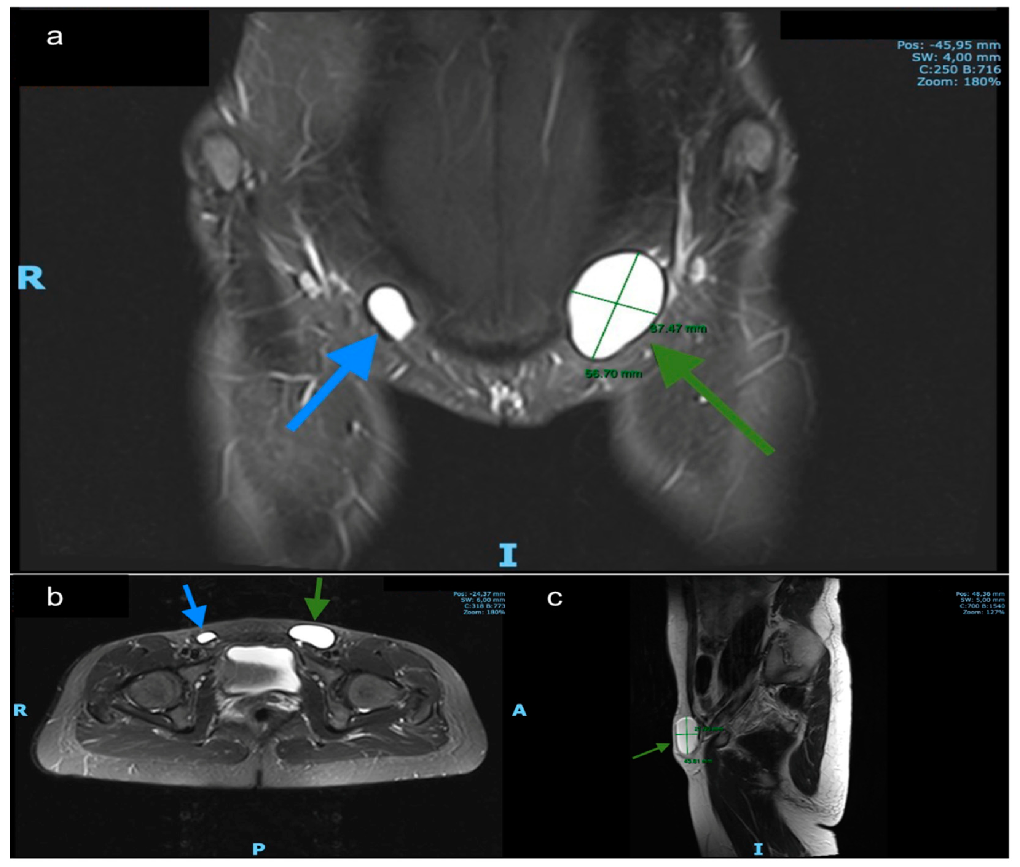
Communicating hydrocele (type 2 cyst of the canal of Nuck) in a 3-year-old girl with intermittent right inguinal bulging. Sagittal US image shows a patent tubular canal of Nuck with fluid content appearing only when standing, which is called a communicating hydrocele (cyst of canal of Nuck type 2). Dashed arrows = internal and external rings of.. patent canal of Nuck is a normal inding dur-ing the 1st year of life but is a predictive anatomic risk factor of hernia. The patent canal of Nuck can have a linear, round, or heart shape (Fig 4). It is usually short in length when there is no her-nia (around 1 cm), and it may or may not contain. small amount of fluid.

Bilateral canal of Nuck cysts; A rare presentation Eurorad

Cyst of Canal of Nuck Jennifer E. Bagley, Mackenzie B. Davis, 2015

Hydrocele of the canal of Nuck presenting as a sausageshaped mass BMJ Case Reports

Canal Of Nuck Cyst Laparoscopic Excision

JCM Free FullText Hydroceles of the Canal of Nuck in Adults—Diagnostic, Treatment and

Bilateral canal of Nuck cysts; A rare presentation Eurorad

Ultrasound picture showing cyst in the canal of Nuck with septae. Download Scientific Diagram

Sonographic appearance of canal of Nuck hydrocele Semantic Scholar

Canal Of Nuck Cyst Laparoscopic Excision YouTube

Cyst of the canal of Nuck ultrasound and MRI appearances Eurorad

Figure 1 from Hydrocele in the Canal of Nuck CT appearance of a developmental groin anomaly

Figure 2 from Hydrocele of the canal of Nuck value of radiological diagnosis. Semantic Scholar

Cyst and Hydrocele of Canal Of Nuck Radiology Shots Short Radiology Cases Round 1 YouTube

Cureus Hydrocele of the Canal of Nuck A Review

Cyst of the Canal of Nuck in adult females A case report and systematic review

Ultrasound picture showing cyst in the canal of Nuck with septae. Download Scientific Diagram

Cyst of Nuck A Disregarded Pathology Journal of Minimally Invasive Gynecology

Cyst of the canal of Nuck ultrasound and MRI appearances Eurorad

Medicina Free FullText The Cyst of the Canal of Nuck Anatomy, Diagnostic and Treatment of

Cyst of Canal of Nuck in a 62YearOld Woman Case Report of a Rare Disease International
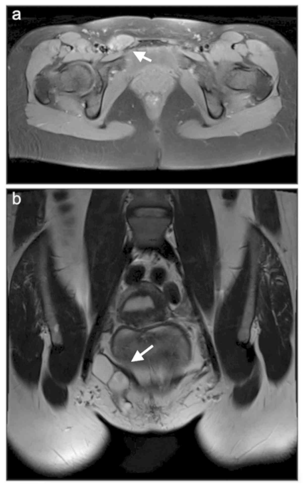
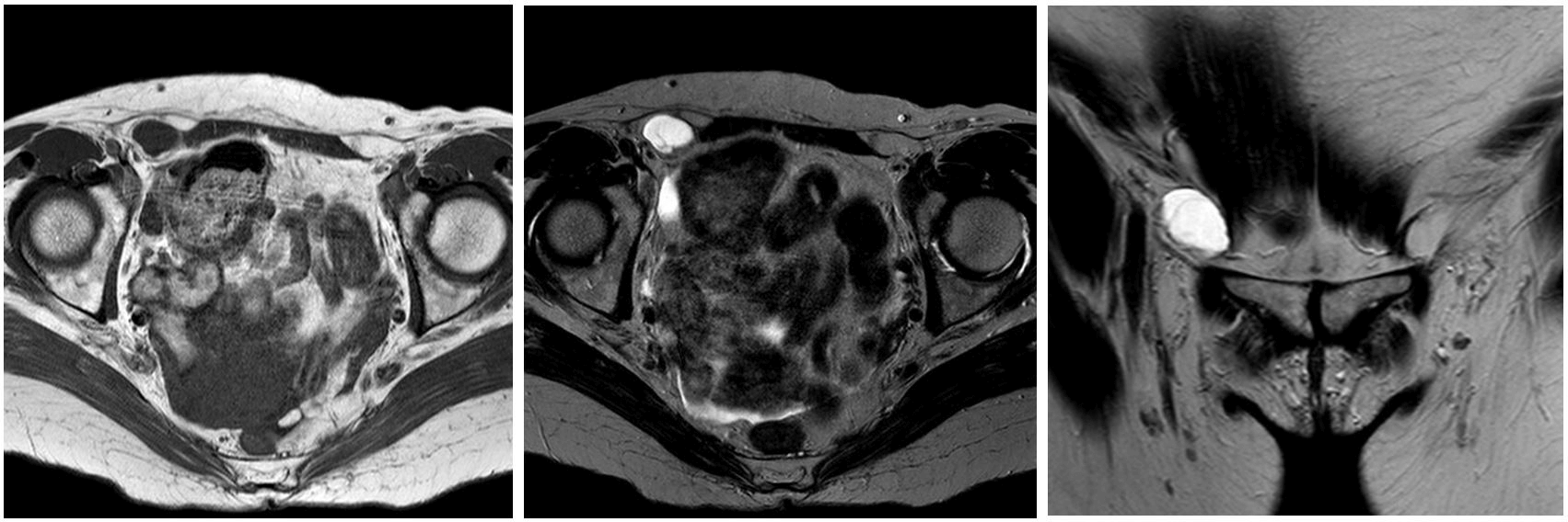
The canal of Nuck is a residue of the peritoneal evagination that runs along the round ligament through the inguinal canal in women. Its partial or total patency can lead to a cystic lymphangioma (CL). CL of the canal of Nuck in an adult female is a rare entity and its clinical diagnosis can be difficult or incorrect.. After interdisciplinary discussion of the external MR imaging by radiologists and surgeons of our institution, a type 1 cyst in the canal of Nuck was diagnosed . The patient decided to proceed with elective surgical therapy. The cyst was excised and a right-sided Lichtenstein hernioplasty was performed to cover the hernia defect.